|
Overview
 A fallen arch is when an arch, even one that has always been flat, has gotten flatter. There are several reasons for this. The bottom of heel tilts toward the inside, making the inside of the ankle appear convex or bowed. The feet often appear to be rotates outward, like a duck. And the arch sags toward the ground. Fallen arches? are more than just flat feet. Having flat feet is not a disease. It is a description of the shape of the foot. Most flat feet have been like that all of the person?s life. Usually they are not painful. Causes Flat feet in adults can appear for a number of reasons. Arches never developed properly. Damage to the tendons that support the arch. Damage or inflammation of the posterior tibial tendon which runs from your lower leg, down and across your ankle, and connects to the middle of the arch. Trauma involving broken bones in the foot. Rheumatoid arthritis. Aging, pregnancy, nerve damage, obesity or even diabetes. Symptoms Most people do not exhibit any symptoms of flat feet, but if the condition is due to an underlying problem, symptoms can include foot pain, mainly in the arch or heel areas, difficulty standing on tiptoes, swelling that occurs on the inside of the ankle, pain in the calf, knee, hip, or lower leg area, both feet lie flat on the ground with no gap, Shoes may not fit properly, heel may tilt away from the midline of the body more than usual, absence of foot arch when standing. If you are experiencing these symptoms and have flat feet, you should consider seeing your doctor or a podiatrist immediately for an examination. Diagnosis You can always give yourself the ?wet test? described above to see whether you have flat feet. Most people who do not notice their flat feet or have no pain associated with them do not think to see a foot doctor. Flat feet can lead to additional problems such as stiffness or pain, however, especially if the condition appears out of nowhere. If you think you may have flat feet, you should seek medical attention to ensure there are no additional issues to worry about. Your doctor will be able to diagnose you with a number of tests. For example, he or she may have you walk around, stand still, or stand on your tiptoes while you are being examined. Your doctor may also examine your foot?s shape and functionality. It?s important to let your foot doctor know about your medical and family history. In some cases, your doctor may order imaging tests such as x-rays or an MRI (magnetic resonance imaging) to determine a cause of your flat foot. If tarsal coalition is suspected in children, a CT scan is often ordered. pes planus treatment Non Surgical Treatment There are home remedies to prevent or manage pain from fallen arches or flat feet. Here are some areas to consider. Wear footwear or shoe inserts that are appropriate to your activity. When pain occurs, try at-home treatment of rest, ice, and over-the-counter nonsteroidal anti-inflammatories, or NSAIDS, such as ibuprofen. Ask your doctor or a physical therapist to show you stretches that can prepare you for feet-intensive activities. Limit or treat risk factors that can make fallen arches or flat feet worse, such as diabetes, high blood pressure, and obesity. Avoid activities that put excessive stress on your feet, such as running on roads. Avoid high-impact sports such as basketball, hockey, soccer, and tennis. Know when to get help. When pain is severe or interferes with activities, it's time to see the doctor for a thorough exam and treatment. Surgical Treatment  Procedures may include the following. Fusing foot or ankle bones together (arthrodesis). Removing bones or bony growths, also called spurs (excision). Cutting or changing the shape of the bone (osteotomy). Cleaning the tendons' protective coverings (synovectomy). Adding tendon from other parts of your body to tendons in your foot to help balance the "pull" of the tendons and form an arch (tendon transfer). Grafting bone to your foot to make the arch rise more naturally (lateral column lengthening).
0 Comments
Overview
Having one leg shorter than the other is a common physical condition. It has two primary causes--structural or functional problems. Structural differences in length can be the result of growth defect, previous injuries or surgeries. Functional differences in length can result from altered mechanics of the feet, knee, hip and/or pelvis. These altered mechanics from functional leg length discrepancy often stem from having an unbalanced foundation.  Causes Some children are born with absence or underdeveloped bones in the lower limbs e.g., congenital hemimelia. Others have a condition called hemihypertrophy that causes one side of the body to grow faster than the other. Sometimes, increased blood flow to one limb (as in a hemangioma or blood vessel tumor) stimulates growth to the limb. In other cases, injury or infection involving the epiphyseal plate (growth plate) of the femur or tibia inhibits or stops altogether the growth of the bone. Fractures healing in an overlapped position, even if the epiphyseal plate is not involved, can also cause limb length discrepancy. Neuromuscular problems like polio can also cause profound discrepancies, but thankfully, uncommon. Lastly, Wilms? tumor of the kidney in a child can cause hypertrophy of the lower limb on the same side. It is therefore important in a young child with hemihypertrophy to have an abdominal ultrasound exam done to rule out Wilms? tumor. It is important to distinguish true leg length discrepancy from apparent leg length discrepancy. Apparent discrepancy is due to an instability of the hip, that allows the proximal femur to migrate proximally, or due to an adduction or abduction contracture of the hip that causes pelvic obliquity, so that one hip is higher than the other. When the patient stands, it gives the impression of leg length discrepancy, when the problem is actually in the hip. Symptoms LLD do not have any pain or discomfort directly associated with the difference of one leg over the other leg. However, LLD will place stress on joints throughout the skeletal structure of the body and create discomfort as a byproduct of the LLD. Just as it is normal for your feet to vary slightly in size, a mild difference in leg length is normal, too. A more pronounced LLD, however, can create abnormalities when walking or running and adversely affect healthy balance and posture. Symptoms include a slight limp. Walking can even become stressful, requiring more effort and energy. Sometimes knee pain, hip pain and lower back pain develop. Foot mechanics are also affected causing a variety of complications in the foot, not the least, over pronating, metatarsalgia, bunions, hammer toes, instep pain, posterior tibial tendonitis, and many more. Diagnosis Leg length discrepancy may be diagnosed during infancy or later in childhood, depending on the cause. Conditions such as hemihypertrophy or hemiatrophy are often diagnosed following standard newborn or infant examinations by a pediatrician, or anatomical asymmetries may be noticed by a child's parents. For young children with hemihypertophy as the cause of their LLD, it is important that they receive an abdominal ultrasound of the kidneys to insure that Wilm's tumor, which can lead to hypertrophy in the leg on the same side, is not present. In older children, LLD is frequently first suspected due to the emergence of a progressive limp, warranting a referral to a pediatric orthopaedic surgeon. The standard workup for LLD is a thorough physical examination, including a series of measurements of the different portions of the lower extremities with the child in various positions, such as sitting and standing. The orthopaedic surgeon will observe the child while walking and performing other simple movements or tasks, such as stepping onto a block. In addition, a number of x-rays of the legs will be taken, so as to make a definitive diagnosis and to assist with identification of the possible etiology (cause) of LLD. Orthopaedic surgeons will compare x-rays of the two legs to the child's age, so as to assess his/her skeletal age and to obtain a baseline for the possibility of excessive growth rate as a cause. A growth chart, which compares leg length to skeletal age, is a simple but essential tool used over time to track the progress of the condition, both before and after treatment. Occasionally, a CT scan or MRI is required to further investigate suspected causes or to get more sophisticated radiological pictures of bone or soft tissue. Non Surgical Treatment Treatment is based on an estimate of how great the difference in leg length will be when the child grows up, Small differences (a half inch or less) do not need treatment. Differences of a half to one inch may require a lift inside the shoe.  how to grow taller exercises Surgical Treatment In growing children, legs can be made equal or nearly equal in length with a relatively simple surgical procedure. This procedure slows down the growth of the longer leg at one or two growth sites. Your physician can tell you how much equalization can be gained by this procedure. The procedure is performed under X-ray control through very small incisions in the knee area. This procedure will not cause an immediate correction in length. Instead, the limb length discrepancy will gradually decrease as the opposite extremity continues to grow and "catch up." Timing of the procedure is critical. The goal is to reach equal leg length by the time growth normally ends. This is usually in the mid-to-late teenage years. Disadvantages of this option include the possibility of slight over-correction or under-correction of the limb length discrepancy. In addition, the patient's adult height will be less than if the shorter leg had been lengthened. Correction of significant limb length discrepancy by this method may make a patient's body look slightly disproportionate because of the shorter leg. In some cases the longer leg can be shortened, but a major shortening may weaken the muscles of the leg. In the thighbone (femur), a maximum of 3 inches can be shortened. In the shinbone, a maximum of 2 inches can be shortened. Overview
 Heel pain is pain in the heel area that can vary in severity and location. It is most common in adults. The heel is the first bone to contact the ground when walking and takes the full force of impact and the resulting shock of bearing weight during motion. Causes There are many reasons why people experience heel pain. Based on what we see in our office, heel pain affects, probably more than one in every four people. A lot of this is caused from conditions within the foot. These conditions could be related to hyper pronation, which is where you get a collapse of the foot or even a high arch of the foot called cavus foot. The underlying cause is something internal, within the bone structure. These problems are usually something you will have all your life. Hyper pronation is a hereditary issue where you can get an under development of a particular bone, usually in the ankle, and it causes a dislocation or a misalignment of the ankle on the heel. It throws off, not only the foot with the bones, joints and ligaments of the foot, but also the bones in the ankle. It affects the internal rotation of the knee, hip, back and causes issues within those areas as well. Hyper pronation is a pretty common, but very under diagnosed condition. Symptoms Plantar fasciitis is a condition of irritation to the plantar fascia, the thick ligament on the bottom of your foot. It classically causes pain and stiffness on the bottom of your heel and feels worse in the morning with the first steps out of bed and also in the beginning of an activity after a period of rest. For instance, after driving a car, people feel pain when they first get out, or runners will feel discomfort for the first few minutes of their run. This occurs because the plantar fascia is not well supplied by blood, which makes this condition slow in healing, and a certain amount of activity is needed to get the area to warm up. Plantar fasciitis can occur for various reasons: use of improper, non-supportive shoes; over-training in sports; lack of flexibility; weight gain; prolonged standing; and, interestingly, prolonged bed rest. Diagnosis A biomechanical exam by your podiatrist will help reveal these abnormalities and in turn resolve the cause of plantar fasciitis. By addressing this cause, the patient can be offered a podiatric long-term solution to his problem. Non Surgical Treatment Anti-inflammatory medication. Heel stretching exercise. Ice application to painful area - twice a day or more, for 10 - 15 minutes. Rest. Supportive, well-fitting, padded shoes. Heel cup, felt pads or orthotics. Night splints - stretches injured fascia and allows healing. Basic treatment. First thing in the morning, before getting out of bed -- massage the bottom of the affected foot or feet for at least five minutes. Ensure that the plantar is stretched and warmed up so that overnight healing remains intact. Before stepping out of bed, be sure that you have soft, padded, supportive shoes or slippers to wear, especially if your flooring is hard, tile or uncarpeted flooring. Surgical Treatment With the advancements in technology and treatments, if you do need to have surgery for the heel, it is very minimal incision that?s done. And the nice thing is your recovery period is short and you should be able to bear weight right after the surgery. This means you can get back to your weekly routine in just a few weeks. Recovery is a lot different than it used to be and a lot of it is because of doing a minimal incision and decreasing trauma to soft tissues, as well as even the bone. So if you need surgery, then your recovery period is pretty quick. no foot pain Prevention  Maintaining flexible and strong muscles in your calves, ankles, and feet can help prevent some types of heel pain. Always stretch and warm-up before exercising. Wear comfortable, properly fitting shoes with good arch support and cushioning. Make sure there is enough room for your toes. Overview
 A bunion is a growth of skin at the joint of the big toe, often a result of enlarged tissue or misaligned bone. In some cases, the bunion may be so extreme that it pushes the big toe inward toward the second toe. Skin and tissue surrounding the joint may experience sensitivity to touch, tenderness and pain. Causes Bunions have a number of causes, primarily genetics and bad choices in footwear. We inherit traits like flat feet, abnormal bone structure, and loose ligaments and tendons from our parents. When our feet are weakened by these traits and we stuff them into high heels or shoes which don?t support our feet correctly, the repeated stress on the front of the foot may contribute to the formation of a bunion. Other contributing factors are jobs that demand a lot of time standing, obesity, and sudden hormonal changes and weight gain, as in pregnancy. Unfortunately, bunions can lead to many other foot conditions as well. The joint behind the big toe carries much of your body weight and when the bunion makes it sore, you shift your weight onto other areas of the foot. That?s why we frequently see crossover toes, overlapping toes, hammer toes, corns, calluses, and ingrown toenails accompanying bunions. As pain in your foot increases, you?ll also reduce your activity, becoming more sedentary, which has its own quality-of-life issues. SymptomsSymptoms of a bunion include irritated skin, sensitivity to touch, and pain when walking or running. Since the bunion may grow so prominent as to affect the shape of the foot, shoes may no longer fit properly, and blisters may form at the site of friction and pressure. Bunions may grow so large that an individual must wear shoes that are a larger size than they would otherwise wear. If the bunion becomes a severe case, walking may become difficult. Diagnosis Your doctor will be able to diagnose a bunion by asking about your symptoms and examining your feet. You may also have blood tests to rule out any other medical conditions, such as rheumatoid arthritis or gout, although this is rare. Your doctor may refer you to a podiatrist or chiropodist (healthcare professionals who specialise in conditions that affect the feet). Non Surgical Treatment Because they are bone deformities, bunions do not resolve by themselves. The goal for bunion treatment is twofold: first, to relieve the pressure and pain caused by irritations, and second to stop any progressive growth of the enlargement. Commonly used methods for reducing pressure and pain caused by bunions include the use of protective padding, often made from felt material, to eliminate the friction against shoes and help alleviate inflammation and skin problems. Removal of corns and calluses on the foot. Changing to carefully fitted footwear designed to accommodate the bunion and not contribute toward its growth. Orthotic devices, both over-the-counter and custom made-to help stabilize the joint and place the foot in the correct position for walking and standing. Exercises to maintain joint mobility and prevent stiffness or arthritis. Splints for nighttime wear to help the toes and joint align properly. This is often recommended for adolescents with bunions, because their bone development may still be adaptable. 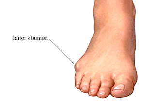 Surgical Treatment Bunion surgery generally involves repositioning the maligned bones with a bone cut (osteotomy) and/or bone mending procedure (fusion). As such, the time it takes for bones to set or mend in the corrected position generally takes six weeks. Smokers and those in poor medical health may take longer to mend the bone. The biology of bone healing is about six weeks, that time frame can't be made quicker. What can be changed is the disability that one experiences while the bone is mending. Prevention If the diagnosis is made early on, such as in preadolescence, bunion development can be slowed and in some cases arrested with the proper supportive shoe gear and custom functional shoe inserts (orthotics). Avoidance of certain athletic activities with improper shoe fit and toe pressure can prevent the symptoms that occur with bunions.
Overview
 A bunion is a deviation and inflammation of joint where the big toe connects to the 1st metatarsal, also known as the 1st MTP. The capsule of the joint is displaced, thickened and enlarged, and the cartilage of the joint is damaged. There are three degrees of bunions: mild, moderate and severe. It is important to know that bunions are not hereditary, although the tendency to overpronate, which is one of the main causes of bunions, has a hereditary component. Patients complain of pain in the joint and have a big toe that points away from the midline of the body. Often, they are only able to wear very wide shoes. Causes Bunions are a result of complex biomechanical changes that occur in your feet. The type of footwear that you wear does cause bunions. We know that foot bunions occur in about 30% of the population of most Western countries but only 3% in Eastern countries. They are seen most commonly in women and become more common as people get older. Tight-fitting shoes are thought to be the main cause of bunions.. Shoes such as high heels and shoes with tight toe boxes (eg womens fashion shoes and cowboy boots) are particularly damaging to the toes. These shoes have a sloping foot bed and a narrow toe box. The slope causes the front of the foot to bear your weight, which encourages your forefoot to widen. Also, the angle pushes your toes into the narrow toe box, causing the toes to become angled and squeezed together. SymptomsThe major symptom of bunions is a hard bump on the outside edge of the foot or at the base of the big toe. Redness, pain and swelling surrounding or at the MTP joint can also occur. Diagnosis A doctor can very often diagnose a bunion by looking at it. A foot x-ray can show an abnormal angle between the big toe and the foot. In some cases, arthritis may also be seen. Non Surgical Treatment Bunion treatment should always start with changing footwear to relieve symptoms and to prevent the bunion from progressing. Shoes with a wide toe-box, minimal slope, and good arch support can help relieve the bunion pain. Some people find that ice application and anti-inflammatory medications can help relieve the inflammation around the bunion. A bunion is a deviation and inflammation of joint where the big toe connects to the 1st metatarsal, also known as the 1st MTP. The capsule of the joint is displaced, thickened and enlarged, and the cartilage of the joint is damaged. There are three degrees of bunions: mild, moderate and severe. It is important to know that bunions are not hereditary, although the tendency to overpronate, which is one of the main causes of bunions, has a hereditary component. Patients complain of pain in the joint and have a big toe that points away from the midline of the body. Often, they are only able to wear very wide shoes. Causes Bunions are a result of complex biomechanical changes that occur in your feet. The type of footwear that you wear does cause bunions. We know that foot bunions occur in about 30% of the population of most Western countries but only 3% in Eastern countries. They are seen most commonly in women and become more common as people get older. Tight-fitting shoes are thought to be the main cause of bunions.. Shoes such as high heels and shoes with tight toe boxes (eg womens fashion shoes and cowboy boots) are particularly damaging to the toes. These shoes have a sloping foot bed and a narrow toe box. The slope causes the front of the foot to bear your weight, which encourages your forefoot to widen. Also, the angle pushes your toes into the narrow toe box, causing the toes to become angled and squeezed together. SymptomsThe major symptom of bunions is a hard bump on the outside edge of the foot or at the base of the big toe. Redness, pain and swelling surrounding or at the MTP joint can also occur. Diagnosis A doctor can very often diagnose a bunion by looking at it. A foot x-ray can show an abnormal angle between the big toe and the foot. In some cases, arthritis may also be seen. Non Surgical Treatment Bunion treatment should always start with changing footwear to relieve symptoms and to prevent the bunion from progressing. Shoes with a wide toe-box, minimal slope, and good arch support can help relieve the bunion pain. Some people find that ice application and anti-inflammatory medications can help relieve the inflammation around the bunion. 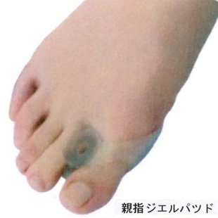 Surgical Treatment If the bunion symptoms does not respond to the conservative measures or if the bunion has progressed past a threshold where these measures are not effective, bunion surgery may be necessary to correct alignment and remove the bunion. A large range of types of surgical procedures for bunions are available and the choice will depend on things like what bone or bones are involved, the angular relationship between the different bones, the amount of damage to the joint and the presence of deformities other than the bunion. Surgical Treatment If the bunion symptoms does not respond to the conservative measures or if the bunion has progressed past a threshold where these measures are not effective, bunion surgery may be necessary to correct alignment and remove the bunion. A large range of types of surgical procedures for bunions are available and the choice will depend on things like what bone or bones are involved, the angular relationship between the different bones, the amount of damage to the joint and the presence of deformities other than the bunion.
|
AuthorWrite something about yourself. No need to be fancy, just an overview. Archives
July 2017
Categories |
 RSS Feed
